by Paula Wolfson, WTOP.com
WASHINGTON – An FDA advisory panel has endorsed the use of a new screening tool for breast cancer.
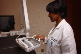
It is a 3-D automated breast ultrasound system which — when combined with conventional mammograms — has been proven to boost cancer detection rates for women with dense breasts.
About 40 percent of American women have dense breast tissue. This highly glandular tissue puts them at higher risk for breast cancer. It also makes it harder for radiologists to read their mammograms when cancer is at its earliest, most treatable stage.
“It can obscure things,” says Dr. Jocelyn Rapelyea, associate director of breast imaging at the George Washington University Medical Faculty Associates.
“Cancer is white. Glandular tissue is white. It makes it very difficult.”
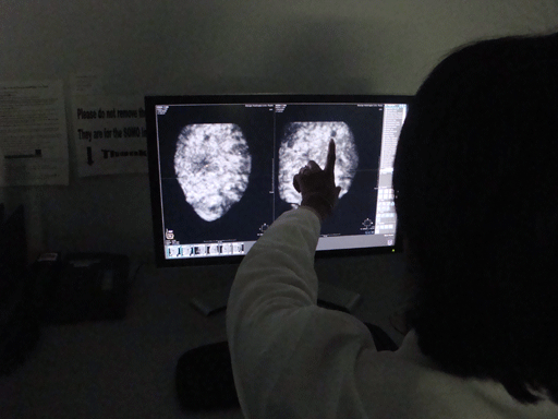
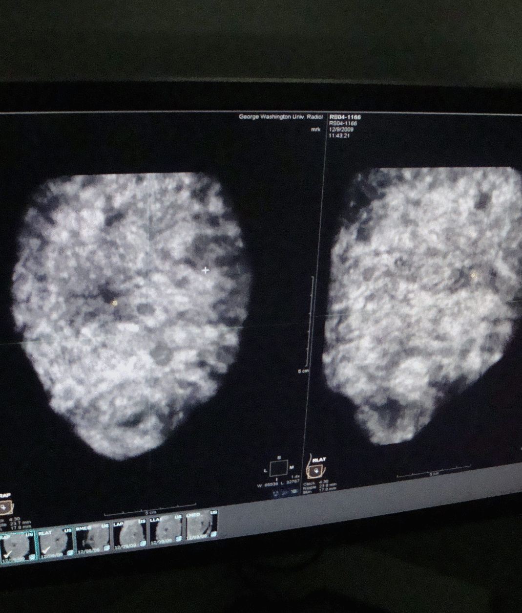
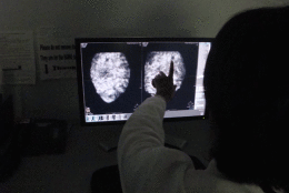
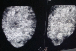
Rapelyea described it like driving through a thick fog and being asked to pick out a single speck. When you add ultrasound to a mammogram, “that lifts that curtain of fog and makes things very ‘contrasty’…cancers pop out as black against that white background,” Rapelyea said.
Ultrasound is already being used as a follow-up to suspicious mammograms. But now, it will be offered as part of the regular screening process for women with dense breast tissue, with a greater likelihood that insurance companies will pick up the tab.
“With the addition of mammography and ultrasound together as a combination in the patient population of dense breast tissue, overall we can increase our detection of breast cancer,” Rapelyea said.
She says the combination has been studied at medical facilities nationwide, including hers. More than 16,000 women have been involved in the clinical trials, with an increased breast cancer detection rate between 24 and 34 percent.
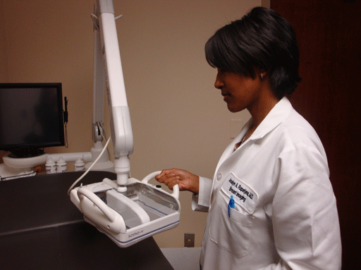
The George Washington University Medical Faculty Associates is currently the only medical center in the region using the Automated Breast Ultrasound System — or ABUS — developed by U-Systems. But Rapelyea predicts FDA approval of the technology for breast cancer screening will prompt others to follow suit.
Unlike mammograms which use radiation, the ABUS uses sound waves to create 3-D images of breast tissue. The exam takes about six minutes per side, and a complete ultrasound/mammogram combination should take no more than half an hour.
Rapelyea cautions ultrasound is not a replacement for a mammogram, but an important add-on for women with dense breast tissue.
“Mammography is still the best tool we have out there for screening,” she said. “It is the only tool that has been shown to reduce mortality.”
Follow WTOP on Twitter.
(Copyright 2012 by WTOP. All Rights Reserved.)


