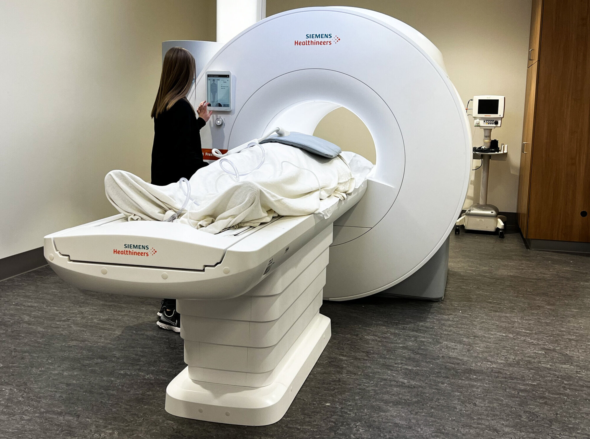
What’s old is new again with an MRI machine that uses a lower magnetic field for patients with implanted defibrillators and pacemakers, and has a wider opening to accommodate people who are severely obese or claustrophobic.
“Over the past 20 years, there has been a real push to get to higher and higher magnetic field strengths because that brings the advantage of a greater signal,” said Dr. Orlando Simonetti, professor of internal medicine and radiology at The Ohio State University College of Medicine.
“But it also brings disadvantages like trying to scan somebody who’s got metal in their body or trying to look at the lungs, and trying to build a magnet that can accommodate bigger patients.”
The metal and wires in implanted devices can distort images on MRI scans using a higher magnetic field.
“It’s kind of interesting that we’ve come full circle with it now, and we’re using our technology and software and algorithm development side to make things work at lower fields,” Simonetti said.
Approved by the Food and Drug Administration last July, the lower magnetic field MRI machine was built by Siemens in collaboration with Simonetti. It has the largest MRI opening to date at 80 centimeters compared to typical machines between 60 and 70 centimeters.
MRI is usually used to scan brains, spines, joints and the abdomen, but it can also be used to create images of the heart and blood vessels.
“The cardiac imaging, it’s going to take a little bit more time, but I would think in the next year to two years, you’ll start to see more and more of this,” Simonetti said. “The problem with low-field MRI is that there is less signal to work with, and we needed to find ways to boost that signal,” Simonetti said.
Ohio State has partnered with Nationwide Children’s Hospital to study use of the new technology with children who have congenital heart disease and need to undergo repeated heart catheterizations. Those catheterizations typically are guided with X-rays that expose patients and doctors doing the procedures to radiation.
“But MRI doesn’t use any X-ray radiation. So that’s very important and an important advantage, especially in children with congenital heart disease … who may get exposed to radiation repeatedly throughout their lives as they go through different surgeries, etc., to help with their condition,” Simonetti said.

“Another advantage of low-field imaging is the potential for lung imaging,” Simonetti said of readings typically taken using nuclear imaging or X-ray CT scans. A mix of air and water in lungs tends to cancel out the MRI signal at higher fields but not as much at lower fields.
“This is an important advancement for patients with cystic fibrosis, pulmonary hypertension, heart failure, COVID-19 and any other disease, where we’re trying to understand the source of shortness of breath and evaluate both the heart and lungs,” Simonetti said.
The new technology using less powerful magnets weighs less and costs significantly less than higher-field machines, and they’re cheaper to install.
“That’s going to be interesting to watch because it’ll open up opportunities in underserved parts of the world, underserved parts of our own country and the ability to put an MRI machine places where maybe that wasn’t possible before,” Simonetti said.
The Ohio State University Wexner Medical Center is the first in the U.S. to install the new full-body lower-magnetic field MRI for patient care.


